Home
/ Ct Pelvis Anatomy Muscles - Atlas Of Ct Anatomy Of The Abdomen W Radiology / There are many muscles that form the pelvic floor, including puborectalis, pubococcygeus, iliococcygeus and coccygeus.
Ct Pelvis Anatomy Muscles - Atlas Of Ct Anatomy Of The Abdomen W Radiology / There are many muscles that form the pelvic floor, including puborectalis, pubococcygeus, iliococcygeus and coccygeus.
Ct Pelvis Anatomy Muscles - Atlas Of Ct Anatomy Of The Abdomen W Radiology / There are many muscles that form the pelvic floor, including puborectalis, pubococcygeus, iliococcygeus and coccygeus.. The gastrocnemius muscle is a complex muscle that is fundamental for walking and posture. The direct (straight) head and indirect (reflected) head. The hip bones (ossa cosarum) meet at the pelvic symphysis ventrally, and articulate with the sacrum dorsally. These four muscles conjoin to attach to the patella as the quadriceps tendon. We study anatomy at the practical anatomy class we study the human body.
The muscles of the pelvis form its floor. This mri male pelvis axial cross sectional anatomy tool is absolutely free to use. Anatomical drawing of the female pelvis. The floor is composed of two muscular layers, the levator ani/coccygeus complex and. • to assess equivocal imaging findings • staging of hepatic neoplasms • metastatic workup of primary malignancies • diagnosis of abdominal masses • assessment of biliary problems • diagnosis of vascular lesions.
Ct Abdomen Anatomy from image.slidesharecdn.com These four muscles conjoin to attach to the patella as the quadriceps tendon. Hepatocellular carcinoma or liver cancer. The direct (straight) head and indirect (reflected) head. This article reviews the anatomical and functional information of the gastrocnemius muscle, its embryological derivation. The rectus femoris has two heads of origin: Ct anatomy of the pelvis. If you want to learn how to read ct scans of the abdomen and pelvis proficiently, this video is an excellent starting point. We'll go through the on this image, we can also see some of the muscles that we talked about specifically the slowest.
ƒ organs and structures of the female pelvis.
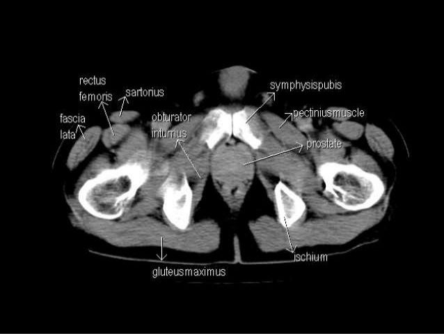
Axial pelvis ct axial femur ct axial femur ct axial knee ct. The muscles of the pelvis, hip and buttock anatomical chart shows how each muscle in this area of the body works with the others, and the various minor systems within the major ones. Hepatocellular carcinoma or liver cancer. The floor is composed of two muscular layers, the levator ani/coccygeus complex and. There are many muscles that form the pelvic floor, including puborectalis, pubococcygeus, iliococcygeus and coccygeus. Pelvic floor muscles that are located wholly within the pelvis. Almost every movement in the body is the outcome of muscle contraction. The muscles are connected with the bones. Learn about anatomy muscles pelvis with free interactive flashcards. The gastrocnemius muscle is a complex muscle that is fundamental for walking and posture. Attached to the bones of the skeletal system are about 700 named. Muscles, connected to bones or internal organs and blood vessels, are in charge for movement. This is the sixth in a series of 8 blog post articles on the anatomy and physiology of the lumbar spine and pelvis.
Muscles, connected to bones or internal organs and blood vessels, are in charge for movement. Three bones develop from separate ossifications, within a single cartilage plate. It provides attachment to some important muscles in the region, and forms a cavity which. Axial pelvis ct axial femur ct axial femur ct axial knee ct. This article reviews the anatomical and functional information of the gastrocnemius muscle, its embryological derivation.
Ct Abdomen And Pelvis Coronal Anatomy In The Male Cute766 from i0.wp.com Ct anatomy of the pelvis. N patient preparation n patient position n scanogram. ƒ organs and structures of the female pelvis. The pelvic girdle consists of two symmetrical halves. The muscles are connected with the bones. The muscular system is responsible for the movement of the human body. They support the pelvic organs especially during increases in intra abdominal pressure and also aid in urinary and faecal. The lateral superficial muscles, the transversus and external and internal oblique muscles, originate on the rib cage and on the pelvis (iliac crest and inguinal ligament) and are attached to the anterior and posterior layers of the sheath of the rectus.
This anatomy section promotes the use of the terminologia anatomica, the international standard of anatomical nomenclature.
Ct anatomy of the pelvis. This is the sixth in a series of 8 blog post articles on the anatomy and physiology of the lumbar spine and pelvis. Although ultrasound is frequently indicated for the primary. This article reviews the anatomical and functional information of the gastrocnemius muscle, its embryological derivation. Pelvic girdle and floor female pelvis and reproductive organs male pelvis and reproductive organs urinary bladder and urethra perineum nerves pelvic organ prolapse kegel exercises. To support the abdominal and pelvic viscera. The direct (straight) head and indirect (reflected) head. N patient preparation n patient position n scanogram. Their main function is contractibility. We study anatomy at the practical anatomy class we study the human body. Use the mouse scroll wheel to move the images up and down alternatively use the tiny arrows (>>) on both side of the image to move the images. Abdominal and pelvic anatomy encompasses the anatomy of all structures of the abdominal and pelvic cavities. Hint you are sitting on it right now.
Axial pelvis ct axial femur ct axial femur ct axial knee ct. The floor is composed of two muscular layers, the levator ani/coccygeus complex and. 0835 lotze anatomy of the pelvic floor. ƒ organs and structures of the female pelvis. There are many muscles that form the pelvic floor, including puborectalis, pubococcygeus, iliococcygeus and coccygeus.
Radiological Anatomy X Ray Ct Mri Kenhub from thumbor.kenhub.com Their main function is contractibility. Axial pelvis ct axial femur ct axial femur ct axial knee ct. Ct anatomy of the pelvis. This mri male pelvis axial cross sectional anatomy tool is absolutely free to use. Rib thorax lumbar pelvis sacrum coccyx femur fibula tibia. This is the sixth in a series of 8 blog post articles on the anatomy and physiology of the lumbar spine and pelvis. The muscles of the pelvis form its floor. Included within the chart are gorgeous illustrations of the pelvic diaphragm, sphincter muscles, gluteus maximus.
Ischial tuberosity which flexor of the knee attaches here?
The direct (straight) head and indirect (reflected) head. ƒ organs and structures of the female pelvis. If you want to learn how to read ct scans of the abdomen and pelvis proficiently, this video is an excellent starting point. Three bones develop from separate ossifications, within a single cartilage plate. 13 what portion of the bony pelvis is the arrow pointing to? Abdominal and pelvic anatomy encompasses the anatomy of all structures of the abdominal and pelvic cavities. Rib thorax lumbar pelvis sacrum coccyx femur fibula tibia. It attaches to the walls of the lesser pelvis, separating the pelvic cavity from the perineum inferiorly (region which includes the in this article, we shall look at the anatomy of the muscles that make up the inferior lining of the cavity; The video covers the most. Online mri & ct sectional anatomy. To maintain the continence of urine and faeces. These and other questions will be addressed as we discuss the gross anatomy and function of the muscles of. They support the pelvic organs especially during increases in intra abdominal pressure and also aid in urinary and faecal.
The hip bones (ossa cosarum) meet at the pelvic symphysis ventrally, and articulate with the sacrum dorsally anatomy muscles pelvis. Muscles, connected to bones or internal organs and blood vessels, are in charge for movement.
